Brainomix Stroke 360 harnesses AI to interpret brain scans in minutes, dramatically improving access to life-saving stroke therapies in 350+ hospitals worldwide.

100%
Increase in mechanical thrombectomy treatment rates¹
64 min
Reduction in door-in-door-out time at primary stroke units
3×
More patients achieving functional independence at 90 days
Calculates ASPECTS score, highlighting regions with hyper- and hypodensities.
Assesses ischemic core based on net water uptake from standard imaging alone — with results equivalent to CT Perfusion and MRI.
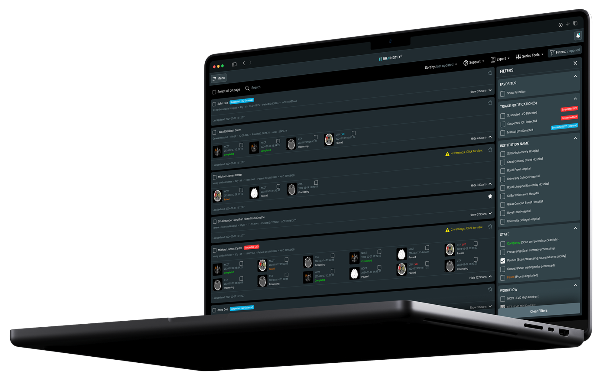
Notifies clinicians of suspected large vessel occlusions and intracranial hemorrhages through real-time smartphone alerts.

Detects large vessel occlusions automatically, flagging suspected LVOs so teams can move faster to thrombectomy.
Highlights vessel density deficit with a color heatmap, quantifying the difference between hemispheres.
Reconstructs vessels in coronal, sagittal and axial views, giving interventionalists a clear picture for treatment planning.

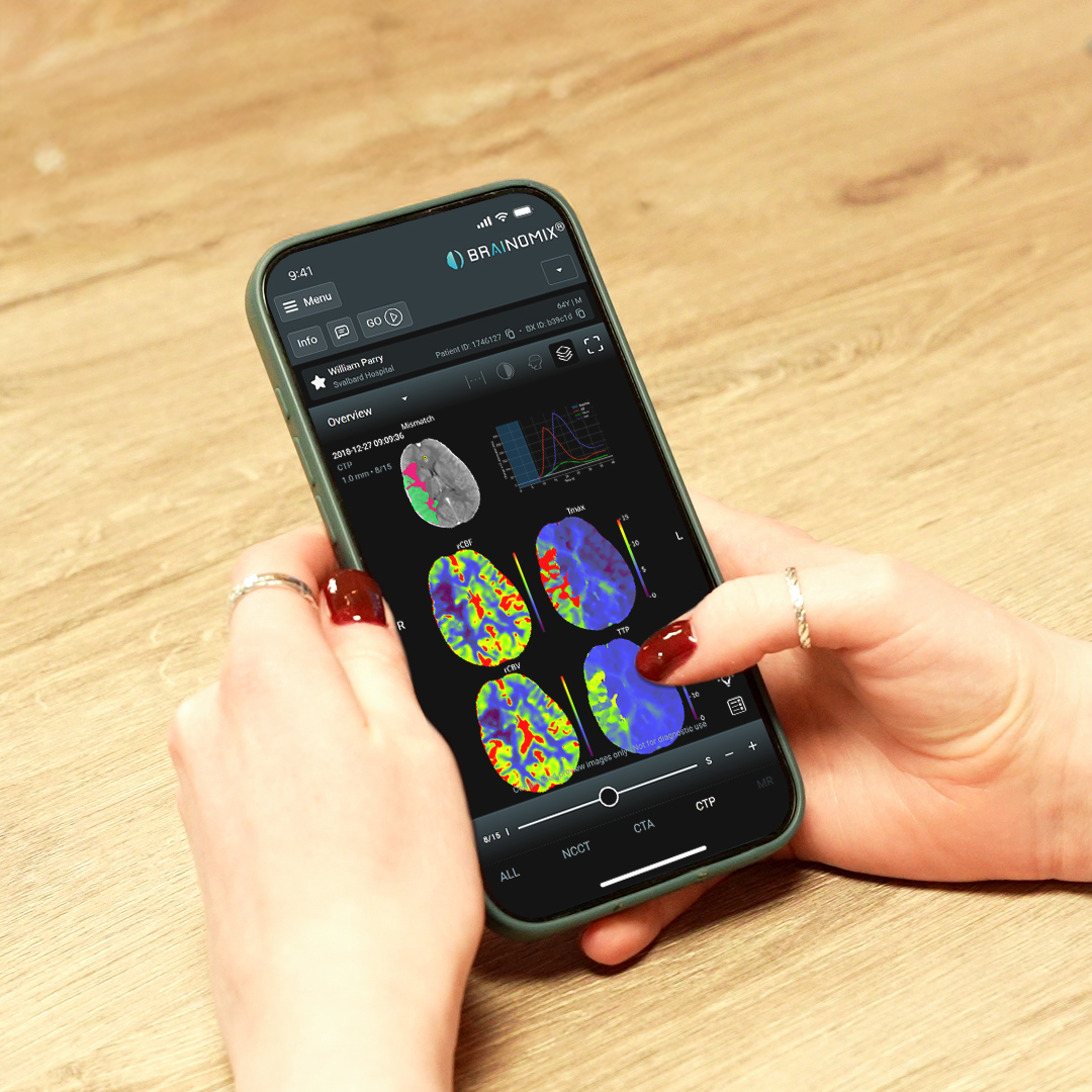
Generates visual reports with metrics mismatch ratio, Tmax >6s, and rCBF <30%, giving clinicians a clear picture of salvageable brain tissue.
Checks patient data against cutting-edge clinical trial criteria to identify more patients who could benefit from EVT therapy.

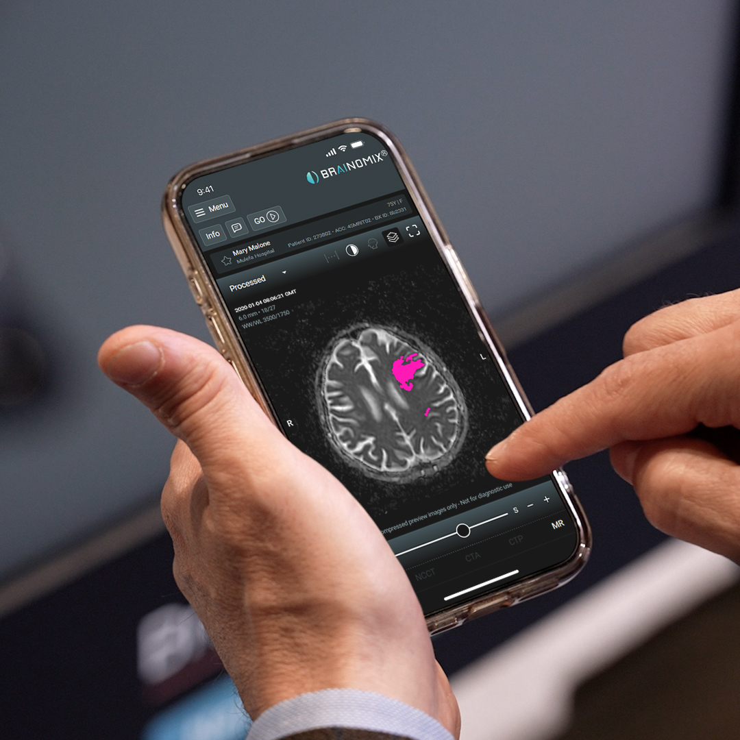
Enables side-by-side visualization of DWI, ADC, b=0, b=max, and perfusion sequences within a single platform to streamline clinician workflows.
Delivers DWI-FLAIR mismatch analysis to help clinicians identify late-window treatment candidates.


University of California,
Los Angeles
Boston Medical Center
West Virginia University
UK’s NHS system
National Deals: Poland, Wales, Hungary, Ireland
Lancet Digital Health study
Mayo Clinic Telestroke Network study
Join 350+ hospitals worldwide already using Brainomix 360 to save more lives, faster.