AI-Powered Stroke Imaging

NCCT

See More. Do More. Save More.

Brainomix 360 Stroke laptop interface
The Most Advanced and Clinically Validated
Stroke AI Platform
Explore Key Features
Smartphone showing Brainomix app with brain CT scan highlighting ischemic stroke regions and volumetric data.
Modality: NCCT
Flags suspected intracranial haemorrhage, measures and outlines
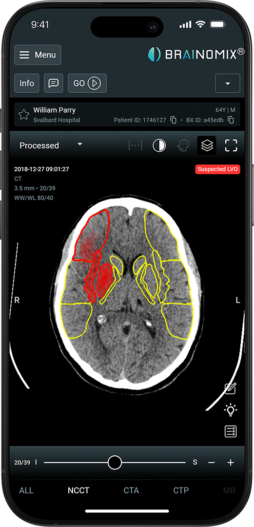
CT brain scan on phone screen showing red and yellow outlines indicating suspected LVO in left brain hemisphere.
Modality: NCCT
Automated quantification of collaterals
CT brain scan on phone screen showing red and yellow outlines indicating suspected LVO in left brain hemisphere.
Modality: NCCT
Automated measure of ischemic core, voxel-by-voxel measurement
CT brain scan on phone screen showing red and yellow outlines indicating suspected LVO in left brain hemisphere.
Modality: NCCT
Provides a standardized quantitative measure of CT hypodensity
Explore Key Features
Mobile screen showing brain vessel CTA scan with a suspected LVO highlighted in red circle on the right side.
Modality: CTA
Detects large vessel occlusions (ICA and M1)
Mobile screen showing brain vessel CTA scan with a suspected LVO highlighted in red circle on the right side.
Modality: CTA
Shows collateral deficit in orange and quantifies.
Brainomix app screen showing CTA results with vessel density 59%, right side occlusion suspected at ICA/Proximal MCA.
Modality: CTA
Automated quantification of collaterals
Smartphone screen showing brain and neck CTA scan with suspected LVO alert and patient details.
Modality: CTA
Axial, coronal, and sagittal views
Axial brain MRI scan showing an area of orange-highlighted abnormality on the right hemisphere.
Modality: NCCT, MRI
Automated measure of edema/swelling
Explore Key Features
Brain imaging scans with color-coded maps and graphs showing cerebral blood flow, volume, and Tmax on phone screen.
Modality: CTP
CBF, Tmax, and more
Brain imaging scans with color-coded maps and graphs showing cerebral blood flow, volume, and Tmax on phone screen.
Modality: CTP
Core and penumbra mismatch
Brain imaging scans with color-coded maps and graphs showing cerebral blood flow, volume, and Tmax on phone screen.
Modality: CTP
Mismatch volume and core volume per side
Brain imaging scans with color-coded maps and graphs showing cerebral blood flow, volume, and Tmax on phone screen.
Modality: CTP
Corrects for patient movement
Brain imaging scans with color-coded maps and graphs showing cerebral blood flow, volume, and Tmax on phone screen.
Modality: CTP
AIF and VOF reference curves
Explore Key Features
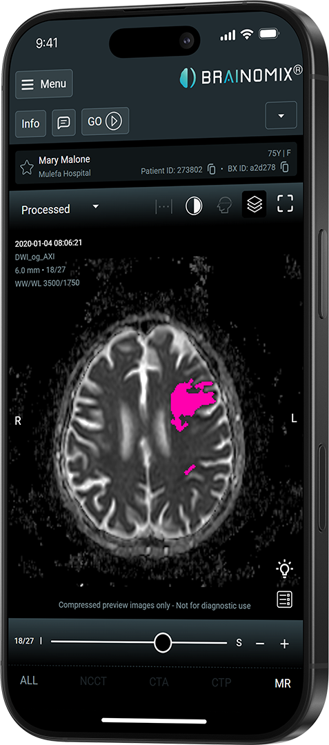
Smartphone display showing brain MRI scan with highlighted pink area indicating abnormality.
Modality: MRI
Maps perfusion mismatch automatically
Smartphone display showing brain MRI scan with highlighted pink area indicating abnormality.
Modality: MRI
Assesses late-window eligibility
Smartphone display showing brain MRI scan with highlighted pink area indicating abnormality.
Modality: MRI
Corrects for patient movement
Smartphone display showing brain MRI scan with highlighted pink area indicating abnormality.
Modality: MRI
Multiple perfusion parameters mapped
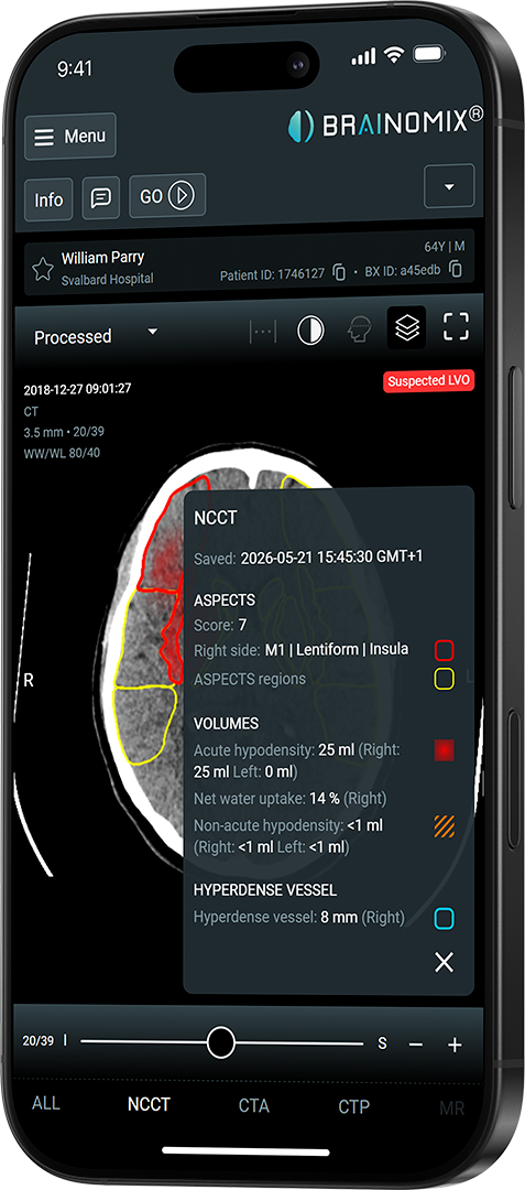
Net Water Uptake
This first-of-its-kind capability provides a standardized, quantitative measure of CT hypodensity. Net water uptake has been associated in the literature with severity of tissue injury and clinical outcomes in acute ischemic stroke.
Read the Evidence
Brainomix app screen shows NCCT results for patient William Parry with ASPECTS score 7, suspected LVO, and volume data.
Real-World Impact

100%

Increase in mechanical thrombectomy treatment rates¹

64 min

Reduction in door-in-door-out time at primary stroke units

More patients achieving functional independence at 90 days

Nagaratnam K, Neuhaus A, Fensome L et al.Artificial intelligence imaging decision support for acute stroke treatment in England: a prospective observational studyThe Lancet Digital Health, 2025; 7

Built to Fit the Way You Work.
01
Scan Acquired
Routine NCCT scan is acquired at your hospital, from there it's sent automatically to Brainomix.
Automated processing
60 seconds
No manual steps
AI-powered analysis
02
Results Delivered
Results are delivered directly to clinicians via PACS, mobile app, email, or web UI within minutes of the scan being acquired.
PACS integration
Mobile app
Email notiication
03
Review and Act
See How We Can Transform Your Stroke Pathway.
Get in Touch
Hands holding a smartphone displaying a brain scan with red and yellow highlighted areas in a medical app.
Unlocking expert-level insights from the scans you already have.

Core volume assessment from NCCT removes the need for advanced imaging in hospitals where CTP or MRI is unavailable.

Validated by leading US stroke centres including Broward Health and UCSF.
Request A Demo
Brain CT scan on phone showing ASPECTS score 7 with red-highlighted affected regions and volume details.
Brainomix 360 Stroke Mobile
Connecting Networks to Facilitate Faster Treatment
The Brainomix 360 Mobile app gives physicians across a stroke network quick, secure access to AI-powered scan results wherever they are. Designed to reduce the workstation bottleneck and keep clinical teams connected when every minute counts.
View AI-processed scan results including pseudonymized patient data from any iOS or Android device, or via web browser.
Learn More
Signal patient eligibility for thrombectomy with a single swipe using the GO button, instantly notifying the relevant clinical team across your network.
Learn More
Coordinate care for each case directly from the app with secure messaging and calling, keeping spoke and hub teams connected.
Learn More
View high-resolution scans with MPR reconstructions in coronal, sagittal, and axial planes to support interventional planning.
Learn More
Phone screen showing Brainomix brain scan analysis with NCCT, CTA, and CTP results for patient William Parry.

Ready to transform stroke care?

Join the hundreds of other hospitals and networks worldwide already using Brainomix 360 Stroke.