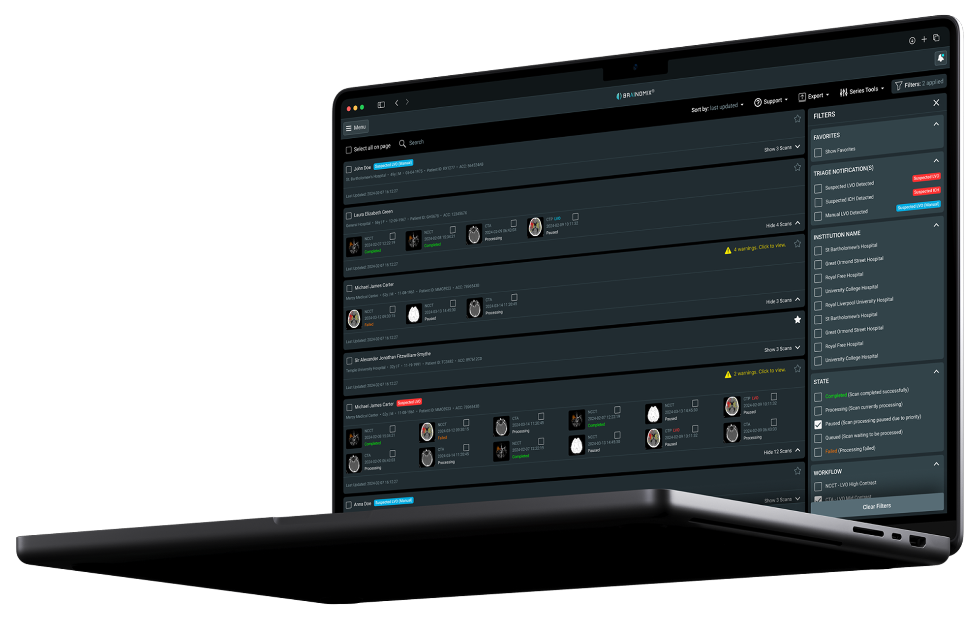
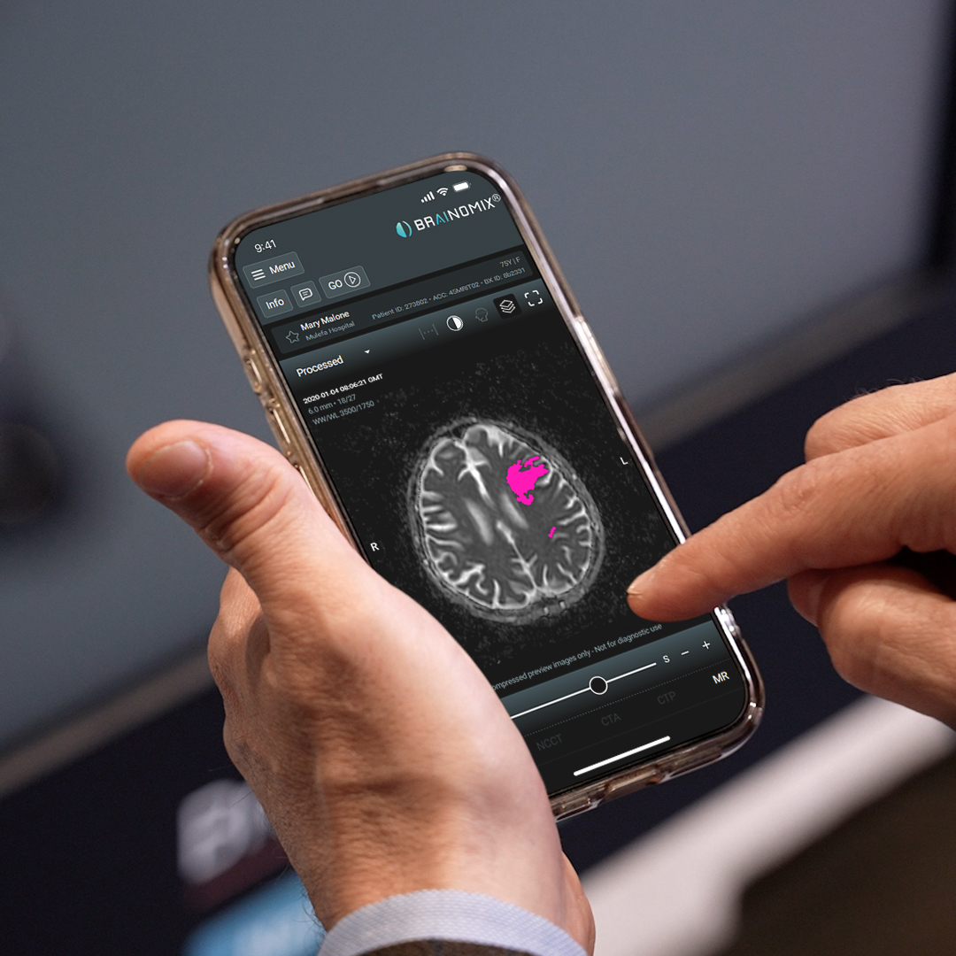
Brainomix Stroke 360 harnesses AI to interpret brain scans in minutes, helping to expand access to life-saving stroke therapies in hundreds of hospitals worldwide.

100%
Increase in mechanical thrombectomy treatment rates¹
64 min
Reduction in door-in-door-out time at primary stroke units
3×
More patients achieving functional independence at 90 days
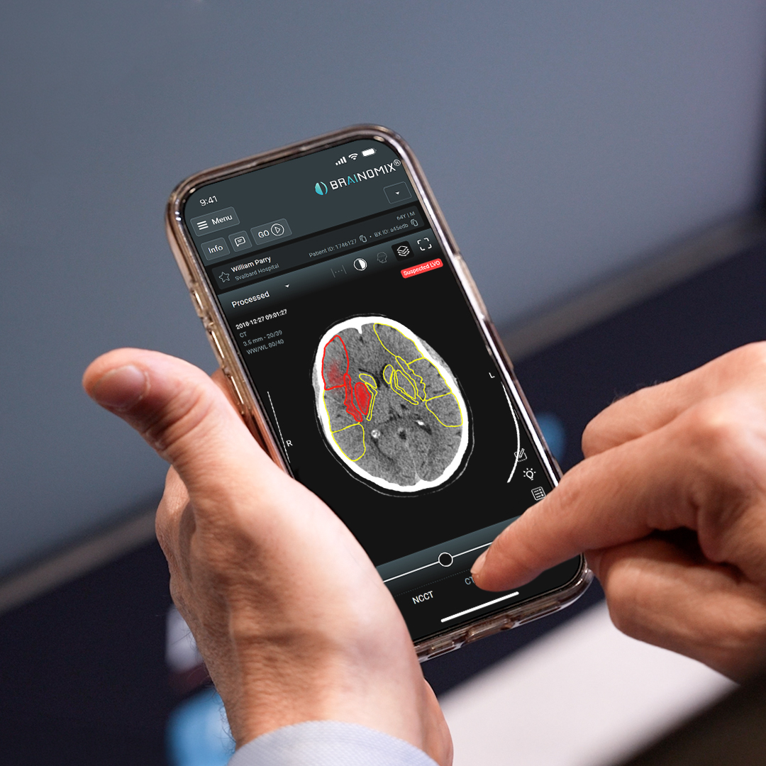
Calculates ASPECTS score, highlighting regions with hyper- and hypodensities.
Calculates net water uptake and assesses ischemic core based on standard imaging alone, with results equivalent to CT Perfusion and MRI.
Notifies clinicians of suspected large vessel occlusions and intracranial hemorrhages through real-time smartphone alerts.

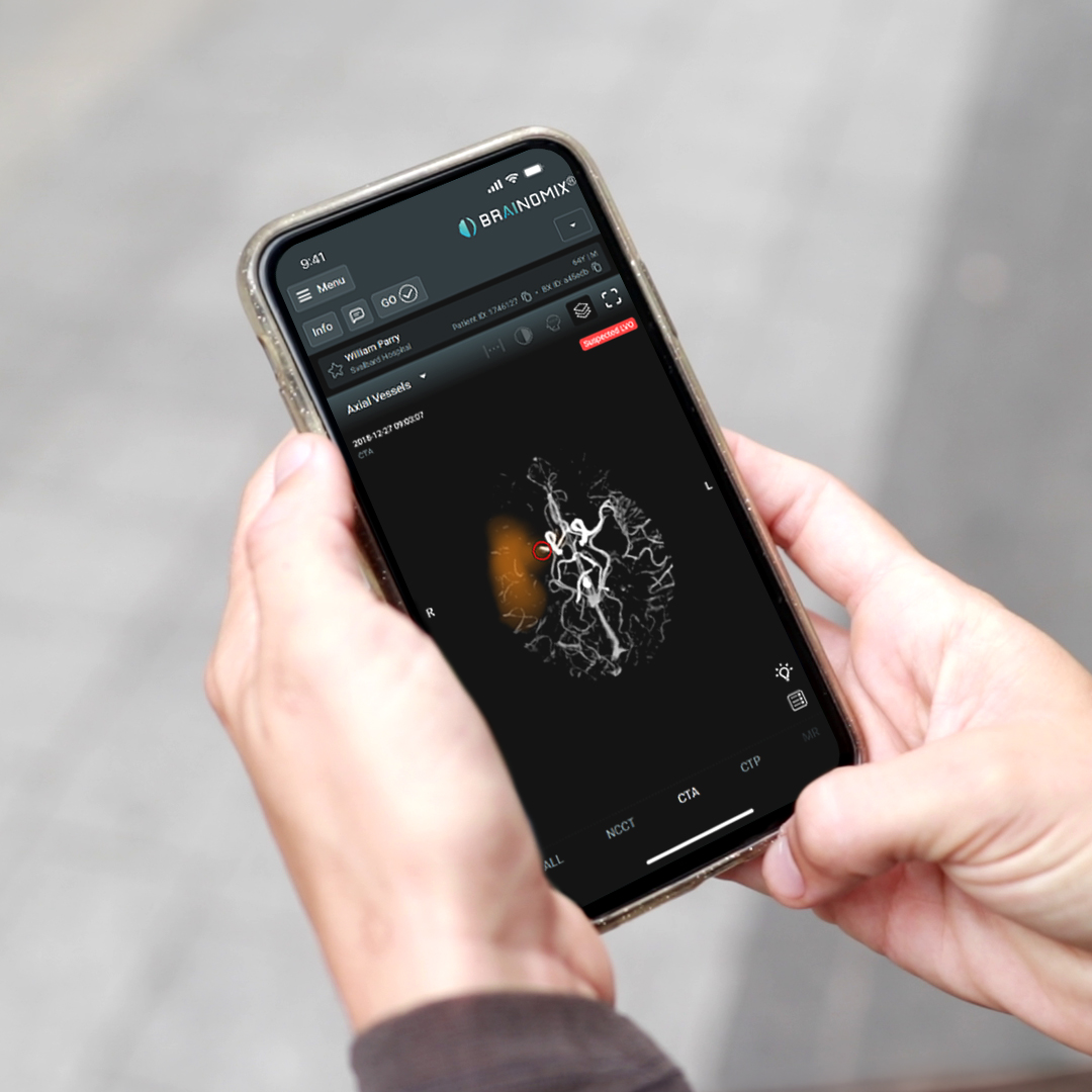
Detects large vessel occlusions automatically, pinpointing the occlusion location so teams can move faster to thrombectomy.
Scores collaterals with the automated CTA-CS score, displayed as a color heatmap comparing blood supply between hemispheres.
Reconstructs vessels in coronal, sagittal and axial views, giving interventionalists a clear picture for treatment planning.

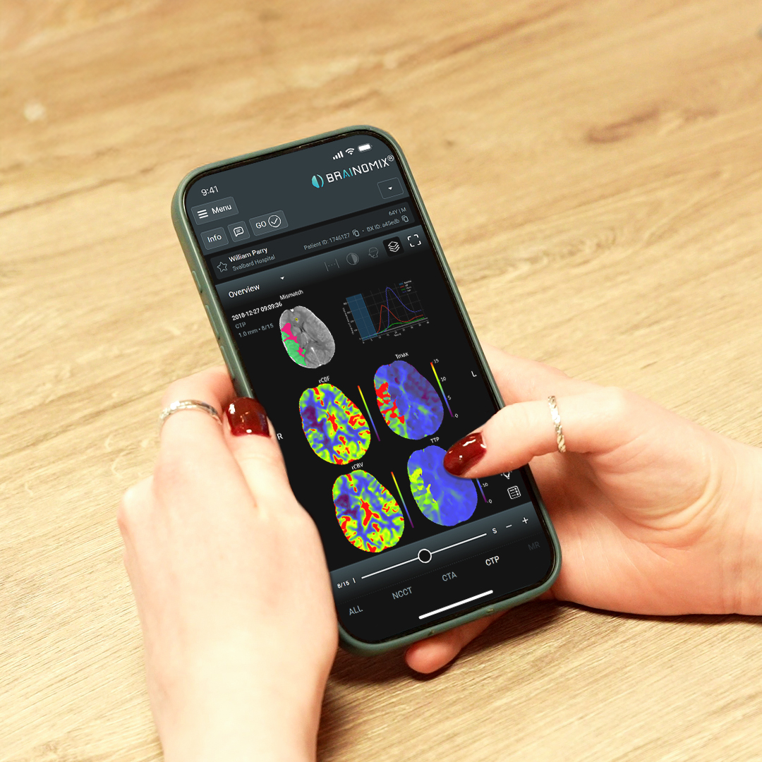
Generates visual reports with metrics mismatch ratio, Tmax >6s, and rCBF <30%, giving clinicians a clear picture of salvageable brain tissue.
Checks patient data against cutting-edge clinical trial criteria to identify more patients who could benefit from EVT therapy.

Enables side-by-side visualization of DWI, ADC, b=0, b=max, and perfusion sequences within a single platform to streamline clinician workflows.
Delivers an automated mismatch map, giving clinicians a clear picture of salvageable tissue.


University of California,
Los Angeles
Boston Medical Center
West Virginia University
UK’s NHS system
National Deals: Poland, Wales, Hungary, Ireland
Lancet Digital Health study
Mayo Clinic Telestroke Network study
Be part of the growing global network of hospitals using Brainomix 360 Stroke.